Onychomycosis means fungal infection of the nail. It is the most common disease of the nails and constitutes about a half of all nail abnormalities.[1]
This condition may affect toenails or fingernails, but toenail infections are particularly common. The prevalence of onychomycosis is about 6-8% in the adult population.[2] Onychomycosis caused by dermatophytes is also known as tinea unguium (tinea of the nails).[3]
Symptoms of Tinea (Onychomycosis)
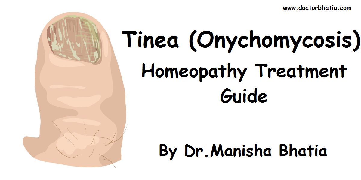
The nail plate can have a thickened, yellow, or cloudy appearance. The nails can become rough and crumbly, or can separate from the nail bed. There is usually no pain or other bodily symptoms, unless the disease is severe. [4]
Dermatophytids are fungus-free skin lesions that sometimes form as a result of a fungus infection in another part of the body. This could take the form of a rash or itch in an area of the body that is not infected with the fungus. Dermatophytids can be thought of as an allergic reaction to the fungus.
Causes of Tinea (Onychomycosis)
Parts of a nail
The causative pathogens of onychomycosis include dermatophytes, Candida, and non-dermatophytic moulds. Dermatophytes are the fungi most commonly responsible for onychomycosis in the temperate western countries; meanwhile, Candida and non-dermatophytic moulds are more frequently involved in the tropics and subtropics with a hot and humid climate.[5]
Trichophyton rubrum is the most common dermatophyte involved in onychomycosis. Other dermatophytes that may be involved are Trichophyton interdigitale, Epidermophyton floccosum, Trichophyton violaceum, Microsporum gypseum, Trichophyton tonsurans, Trichophyton soudanense (considered by some to be an African variant of T. rubrum rather than a full-fledged separate species) and the cattle ringworm fungus Trichophyton verrucosum. A common outdated name that may still be reported by medical laboratories is Trichophyton mentagrophytes for T. interdigitale. The name T. mentagrophytes is now restricted to the agent of favus skin infection of the mouse; though this fungus may be transmitted from mice and their danders to humans, it generally infects skin and not nails.
Other causative pathogens include Candida and non-dermatophytic moulds, in particular members of the mould genera Scytalidium (name recently changed to Neoscytalidium), Scopulariopsis, and Aspergillus. Candida mainly cause fingernail onychomycosis in people whose hands are often submerged in water. Scytalidium mainly affects people in the tropics, though it persists if they later move to areas of temperate climate.
Other moulds more commonly affect people older than 60 years, and their presence in the nail reflects a slight weakening in the nail’s ability to defend itself against fungal invasion.
Classification of Tinea (Onychomycosis)
- Distal Subungual Onychomycosis
- The most common form of tinea unguium usually caused by Trichophyton rubrum, which invades the nail bed and the underside of the nail plate.
- White Superficial Onychomycosis
- Caused by fungal invasion of the superficial layers of the nail plate to form “white islands” on the plate. Accounts for only 10 percent of onychomycosis cases.
- Proximal Subungual Onychomycosis
- Fungal penetration of the newly formed nail plate through the proximal nail fold. It is the least common form of tinea unguium in healthy people but found more commonly when the patient is immunocompromised.
- Candidal Onychomycosis
- Candida species invade fingernails usually occurring in persons who frequently immerse their hands in water. This normally requires the prior damage of the nail by infection or trauma.
- Total Dystrophic Onychomycosis
- Total destruction of the nail plate. It is the end result of any of the above four types.
Diagnosis for Tinea (Onychomycosis)
If all nails are affected then fungal infection is improbable. To avoid misdiagnosis as psoriasis, lichen planus, contact dermatitis, trauma, nail bed tumor or yellow nail syndrome, laboratory confirmation may be necessary. The three main approaches are potassium hydroxide smear, culture and histology. This involves microscopic examination and culture of nail scrapings or clippings. Recent results indicate that the most sensitive diagnostic approaches are direct smear combined with histological examination, [6] and nail plate biopsy using periodic acid-Schiff stain.[7]
Treatment of Tinea (Onychomycosis)
Onychomycosis due to Trychophyton rubrum, right and left great toe.
Treatment of onychomycosis is challenging because the infection is embedded within the nail and is difficult to reach. As a result full removal of symptoms is very slow and may take a year or more.
Pharmacologic Rx
Most treatments are either systemic antifungal medications such as terbinafine and itraconazole, or topical such as nail paints containing ciclopirox or amorolfine. There is also evidence for combining systemic and topical treatments.[8]
For superficial white onychomycosis systemic rather than topical antifungal therapy is advised.[9]
Relative effectiveness of treatments
In July 2007 a meta-study reported on clinical trials for topical treatments of fungal nail infections. The study included 6 randomised controlled trials dating up to March 2005.[10] The main findings are:
- There is some evidence that ciclopiroxolamine and butenafine are both effective but both need to be applied daily for prolonged periods (at least 1 year).
- There is evidence that topical ciclopiroxolamine has poor cure rates and that amorolfine might be substantially more effective.
- Further research into the effectiveness of antifungal agents for nail infections is required.
A 2002 study compared the efficacy and safety of terbinafine in comparison with placebo, itraconazole and griseofulvin in treating fungal infections of the nails.[11] The main findings were that for reduced fungus terbinafine was found to be significantly better than itraconazole and griseofulvin, and terbinafine was better tolerated than itraconazole.
- A small study in 2004 showed that ciclopirox nail paint was more effective when combined with topical urea cream.[12]
- A study of 504 patients in 2007 found that aggressive debridement of the nail combined with oral terbinafine significantly reduced symptom frequency over terbinafine alone.[13]
- A 2007 randomised clinical trial with 249 patients show that a combination of amorolfine nail lacquer and oral terbinafine enhances clinical efficacy and is more cost-effective than terbinafine alone.[14]
Drug pipeline
Most drug development activities are focused on
- the discovery of new antifungals
- novel delivery methods to promote access of existing antifungal drugs into the infected nail plate
Active clinical trials investigating Onychomycosis:[15]
Phase III
- A medicinal nail lacquer, NM100060 from NexMed,[16] contains terbinafine as the active ingredient and a permeation enhancer which facilitates the delivery of the drug into the nail bed where the fungus resides. Commercial sale of the product is expected to begin no earlier than in 2008.[17]
- A comparison of delivery methods for Itraconzole.[18]
- Safety and tolerability of topical Terbinafine.[19]
Phase II
- A topical treatment, AN-2690, is being developed by Schering-Plough Corp. It is active against Trichophyton species.[20]
- Posaconazole, taken orally.[21]
A non-pharmalogical approach with encouraging preliminary results is ultraviolet germicidal irradiation which has been shown to deactivate dematophytes both in vitro and ex vivo.[22]
Natural remedies
As with many diseases, there are also some scientifically unverified folk or alternative medicine remedies.
- Australian tea tree oil.[23][24] There is insufficient information to make recommendations for or against the use of tea tree oil for onychomycosis.[25]
- Grapefruit seed extract as a natural antimicrobial is not demonstrated. Its effectiveness is scientifically unverified. Multiple studies indicate that the universal antimicrobial activity is due to contamination with synthetic preservatives that were unlikely to be made from the seeds of the grapefruit.[26][27][28][29][30]
Prevention for Tinea (Onychomycosis)
- Avoid walking barefoot in public areas such as showers, communal changing rooms.
- Keeping feet clean and dry.
- Avoid sharing shoes and socks
Homeopathy Treatment for Tinea (Onychomycosis)
Keywords: homeopathy, homeopathic, treatment, cure, remedy, remedies, medicine
Homeopathy treats the person as a whole. It means that homeopathic treatment focuses on the patient as a person, as well as his pathological condition. The homeopathic medicines are selected after a full individualizing examination and case-analysis, which includes the medical history of the patient, physical and mental constitution, family history, presenting symptoms, underlying pathology, possible causative factors etc. A miasmatic tendency (predisposition/susceptibility) is also often taken into account for the treatment of chronic conditions. A homeopathy doctor tries to treat more than just the presenting symptoms. The focus is usually on what caused the disease condition? Why ‘this patient’ is sick ‘this way’. The disease diagnosis is important but in homeopathy, the cause of disease is not just probed to the level of bacteria and viruses. Other factors like mental, emotional and physical stress that could predispose a person to illness are also looked for. No a days, even modern medicine also considers a large number of diseases as psychosomatic. The correct homeopathy remedy tries to correct this disease predisposition. The focus is not on curing the disease but to cure the person who is sick, to restore the health. If a disease pathology is not very advanced, homeopathy remedies do give a hope for cure but even in incurable cases, the quality of life can be greatly improved with homeopathic medicines.
The homeopathic remedies (medicines) given below indicate the therapeutic affinity but this is not a complete and definite guide to the homeopathy treatment of this condition. The symptoms listed against each homeopathic remedy may not be directly related to this disease because in homeopathy general symptoms and constitutional indications are also taken into account for selecting a remedy. To study any of the following remedies in more detail, please visit the Materia Medica section at Hpathy.
None of these medicines should be taken without professional advice and guidance.
Homeopathy Remedies for Tinea (Onychomycosis) :
Ail., arg-n., calc., carb-an., carb-s., carb-v., chin., cina., cupr., cupr-ac., cur., fil., form., frag-v., gran., graph., grat., kali-c., kali-i., mag-m., merc., nat-c., nat-s., nux-v., petr., phos., plat., puls., sabad., sant., sep., sil., stann., sulph., ter., thuj., valer.
References
- ^ Szepietowski JC, Salomon J (2007). “Do fungi play a role in psoriatic nails?”. Mycoses 50 (6): 437–42. doi:. PMID 17944702.
- ^ “Impact 07 – Dermatology” (PDF). Bay Bio (2007). Retrieved on 2007-06-13.
- ^ Perea S, Ramos MJ, Garau M, Gonzalez A, Noriega AR, del Palacio A (2000). “Prevalence and risk factors of tinea unguium and tinea pedis in the general population in Spain“. J. Clin. Microbiol. 38 (9): 3226–30. PMID 10970362.
- ^ Onychomycosis at eMedicine
- ^ Chi CC, Wang SH, Chou MC (2005). “The causative pathogens of onychomycosis in southern Taiwan”. Mycoses 48 (6): 413–20. doi:. PMID 16262878.
- ^ Karimzadegan-Nia M, Mir-Amin-Mohammadi A, Bouzari N, Firooz A (2007). “Comparison of direct smear, culture and histology for the diagnosis of onychomycosis”. Australas. J. Dermatol. 48 (1): 18–21. doi:. PMID 17222296.
- ^ Weinberg JM, Koestenblatt EK, Tutrone WD, Tishler HR, Najarian L (2003). “Comparison of diagnostic methods in the evaluation of onychomycosis”. J. Am. Acad. Dermatol. 49 (2): 193–7. PMID 12894064.
- ^ Rodgers P, Bassler M (2001). “Treating onychomycosis“. Am Fam Physician 63 (4): 663–72, 677–8. PMID 11237081.
- ^ Baran R, Faergemann J, Hay RJ (2007). “Superficial white onychomycosis–a syndrome with different fungal causes and paths of infection”. J. Am. Acad. Dermatol. 57 (5): 879–82. doi:. PMID 17610995.
- ^ Crawford F, Hollis S (2007). “Topical treatments for fungal infections of the skin and nails of the foot”. Cochrane Database Syst Rev (3): CD001434. doi:. PMID 17636672.
- ^ Haugh M, Helou S, Boissel JP, Cribier BJ (2002). “Terbinafine in fungal infections of the nails: a meta-analysis of randomized clinical trials”. Br. J. Dermatol. 147 (1): 118–21. PMID 12100193.
- ^ Mitchel L. Zoler (April 1, 2004). “Boosts drug entry into nails: urea, ciclopirox combo tested for onychomycosis.(Focus on Skin Disorders)”, Internal Medical News, pp. 69.
- ^ Potter LP, Mathias SD, Raut M, Kianifard F, Landsman A, Tavakkol A (2007). “The impact of aggressive debridement used as an adjunct therapy with terbinafine on perceptions of patients undergoing treatment for toenail onychomycosis”. The Journal of dermatological treatment 18 (1): 46–52. PMID 17373090.
- ^ Baran R, Sigurgeirsson B, Berker DD, et al (2007). “A multicentre, randomized, controlled study of the efficacy, safety and cost-effectiveness of a combination therapy with amorolfine nail lacquer and oral terbinafine compared with oral terbinafine alone for the treatment of onychomycosis with matrix involvement”. British Journal of Dermatology 157: 149. doi:. PMID 17553051.
- ^ clinicaltrials.gov
- ^ ClinicalTrials.gov NCT00459537
- ^ NEXMED Medicines of the Future
- ^ ClinicalTrials.gov NCT00356915
- ^ ClinicalTrials.gov NCT00443820 and ClinicalTrials.gov NCT00443898
- ^ Barak O, Loo DS (2007). “AN-2690, a novel antifungal for the topical treatment of onychomycosis”. Curr Opin Investig Drugs 8 (8): 662–8. PMID 17668368.
- ^ ClinicalTrials.gov NCT00491764
- ^ PubMed
- ^ Buck DS, Nidorf DM, Addino JG (1994). “Comparison of two topical preparations for the treatment of onychomycosis: Melaleuca alternifolia (tea tree) oil and clotrimazole”. J Fam Pract 38 (6): 601–5. PMID 8195735.
- ^ Nenoff P, Haustein UF, Brandt W (1996). “Antifungal activity of the essential oil of Melaleuca alternifolia (tea tree oil) against pathogenic fungi in vitro”. Skin Pharmacol. 9 (6): 388–94. PMID 9055360.
- ^ “Tea tree oil (Melaleuca alternifolia)“. Drugs & Supplements. Mayo Clinic (May 1, 2006). Retrieved on 2008-01-29.
- ^ von Woedtke T, Schlüter B, Pflegel P, Lindequist U, Jülich WD (1999). “Aspects of the antimicrobial efficacy of grapefruit seed extract and its relation to preservative substances contained”. Pharmazie 54 (6): 452–6. PMID 10399191.
- ^ Sakamoto S, Sato K, Maitani T, Yamada T (1996). “[Analysis of components in natural food additive “grapefruit seed extract” by HPLC and LC/MS]” (in Japanese). Eisei Shikenjo h?koku. Bulletin of National Institute of Hygienic Sciences (114): 38–42. PMID 9037863.
- ^ Takeoka G, Dao L, Wong RY, Lundin R, Mahoney N (2001). “Identification of benzethonium chloride in commercial grapefruit seed extracts”. J. Agric. Food Chem. 49 (7): 3316–20. PMID 11453769.
- ^ Takeoka GR, Dao LT, Wong RY, Harden LA (2005). “Identification of benzalkonium chloride in commercial grapefruit seed extracts”. J. Agric. Food Chem. 53 (19): 7630–6. doi:. PMID 16159196.
- ^ Ganzera M, Aberham A, Stuppner H (2006). “Development and validation of an HPLC/UV/MS method for simultaneous determination of 18 preservatives in grapefruit seed extract”. J. Agric. Food Chem. 54 (11): 3768–72. doi:. PMID 16719494.